A Closer Look at the Heart
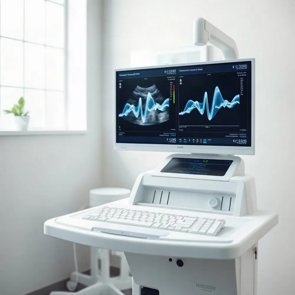
An echocardiogram, often called an echo or heart ultrasound, uses sound waves to create moving pictures of a child’s heart and nearby blood vessels. Unlike an X-ray, it does not use radiation, and a standard echo is noninvasive and generally painless. A child may feel light pressure as the ultrasound probe moves across the chest, but the scan does not involve needles.
At Federal Way Pediatrics, an echocardiogram can help a pediatric cardiology clinician assess the heart’s structure and blood flow. Pediatric echocardiography can also show how the heart muscle is working. The following sections explain why a clinician may recommend an echo, what children can expect during the scan, and how families can prepare.
What an Echo Can Show
 What an Echo Can Show
A pediatric echocardiogram provides moving images clinicians use to assess the heart’s structure and function. The views can show the chambers and walls, how the valves open and close, how strongly the heart pumps, and the direction of blood flow. Pediatric echocardiography can also help clinicians measure blood-flow speed and estimate pressure inside the heart.
Images may reveal openings between chambers, unusual valve shape or movement, narrowed valves, fluid around the heart, or changes in chamber size and wall thickness. They can also show altered blood flow or pumping function. These findings help clinicians assess concerns such as a heart murmur or symptoms, but an image is only one part of the evaluation.
A cardiologist interprets the images alongside your child’s medical history, symptoms, and examination. Needing an echo does not, on its own, mean that a serious heart problem has been found. The test may be recommended to investigate a concern, check another finding, or monitor a known condition; the care team can explain what prompted it and whether any follow-up is needed. At federalwaypediatrics.com, pediatric cardiology care includes discussing what the test is meant to assess and what the results may mean for your child.
A fetal echocardiogram is a focused ultrasound examination of an unborn baby’s heart. A sonographer applies gel to the pregnant patient’s abdomen and moves a handheld probe over the skin to obtain images. This noninvasive test uses sound waves, not radiation, and a pediatric cardiologist reviews the images to assess the developing heart.
When a Clinician May Recommend One
A clinician may recommend an echocardiogram when an exam or medical history raises a question about a child’s heart. Common reasons include a heart murmur, a known or suspected congenital heart condition, or a concern noticed during pregnancy or after birth. An echo lets clinicians assess the heart’s structure and function, including its walls, valves, blood vessels, and pumping strength, as described by KidsHealth.
Symptoms that may prompt an assessment include unusual shortness of breath, fainting, dizziness, chest discomfort, or tiring sooner than expected during activity. For babies, tiring, sweating, or breathing quickly during feeds may also be worth discussing. A family history of certain heart problems can be relevant, too. These signs do not automatically mean a child needs imaging. Many murmurs and occasional skipped beats are harmless, and the clinician decides whether an echo or another step is appropriate.
If you have concerns, a pediatric cardiology evaluation at Federal Way Pediatrics can help clarify whether further assessment is needed. You can also read about reasons an echo may be recommended. Seek urgent medical care for severe difficulty breathing, fainting during exercise, or symptoms that appear suddenly or are getting worse.
What Happens During the Scan
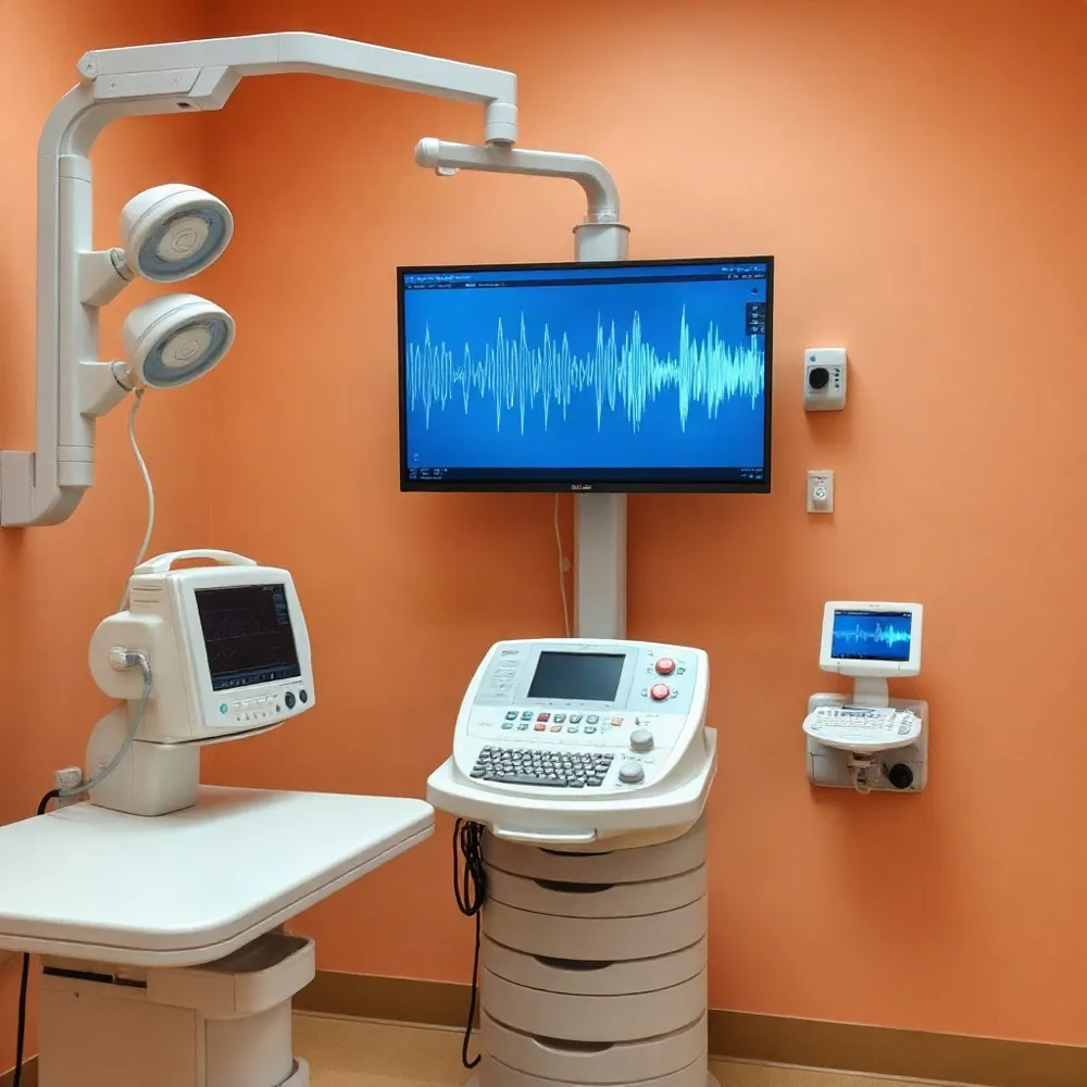 A standard transthoracic echocardiogram is a noninvasive ultrasound scan. Your child usually lies on an exam table, with clothing moved or removed from the waist up. Small electrodes may be placed on the chest to monitor the heartbeat while a sonographer applies gel and moves a handheld transducer over the chest and sometimes the abdomen to capture images. The scan uses sound waves rather than radiation.
The gel may feel cool or warm, and your child may notice gentle pressure from the transducer. The scan itself is generally painless. Staying as still as possible helps the sonographer collect clear images, but the team may allow breaks if your child needs to move or settle. A caregiver can often stay nearby for reassurance. A familiar toy, book, or video may also help; Children’s Wisconsin offers practical suggestions for preparing a child.
The imaging portion commonly takes about 30 to 60 minutes, depending on the images needed and how easily your child can remain still. Allow extra time for check-in and getting ready. For more detail about the visit, see what to expect during an echocardiogram.
At federalwaypediatrics.com, pediatric echocardiograms are part of the practice’s pediatric cardiology services. If a different type of echo or sedation is planned, the procedure and preparation may differ, so ask your child’s care team what to expect and follow its specific instructions.
Preparing Your Child and Getting Results
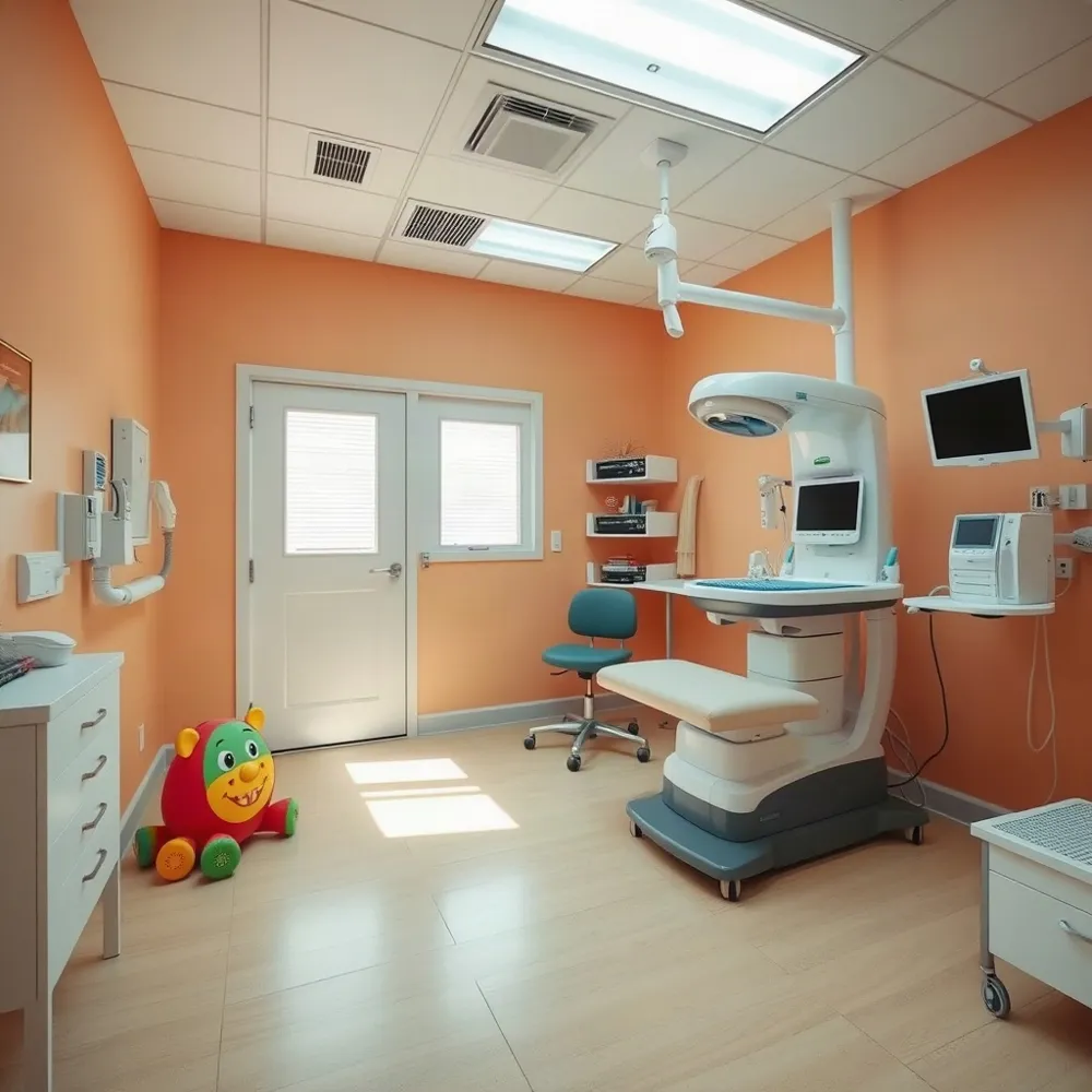 For a standard echocardiogram, most children can eat, drink, and take their usual medicines. Fasting is generally not needed, but follow your care team’s instructions, especially if sedation or a different type of study is planned. KidsHealth’s echocardiogram guide also advises families to check with their clinician about preparation.
Choose comfortable clothing that is easy to remove, and bring a favorite toy, book, or other distraction. A simple explanation can help: the clinician will put gel on the chest and move a small probe over the skin to take pictures. The gel may feel cool, and your child may be asked to lie still or change position. Parents or caregivers can usually stay nearby to offer reassurance. For more practical ideas, see this guide to preparing a child for an echocardiogram.
Bring a list of your child’s medicines and any questions or concerns. Tell the care team about special healthcare needs, difficulty lying still, or worries about the test. If sedation or another kind of echocardiogram is planned, ask the clinic directly about food, drink, and medication instructions. Do not apply fasting rules from another hospital to your child’s appointment.
A trained sonographer typically obtains the images, and a pediatric cardiologist interprets them alongside your child’s symptoms, examination, and medical history. The cardiologist may review images during the visit or request additional views. The care team will explain whether findings are reassuring and whether follow-up, further testing, or treatment is recommended.
Result timing and how families receive updates vary by clinic. Ask when to expect a discussion and how results will be shared. If anything is unclear afterward, contact your child’s care team for an explanation.
Costs and Practical Questions
There is no single price for a pediatric echocardiogram. What a family pays can vary by provider, facility, location, and insurance plan. Hospital price listings may show wide differences, but they do not reliably predict a child’s out-of-pocket cost or a local clinic’s rate.
Coverage may depend on whether the test is considered medically necessary, whether the clinician and facility are in network, and whether the plan requires a referral or prior authorization. A deductible, copay, or coinsurance may also affect the final bill. Families comparing coverage can review how insurance terms affect costs.
Before scheduling, contact both your insurer and the cardiology office to confirm coverage and request an estimate. Ask whether the estimate includes separate facility or interpretation charges, and check whether authorization must be obtained before the appointment.
Questions Help Families Feel Prepared
Before the appointment, families can ask why the echocardiogram was recommended, what preparation applies to their child, and when and how results will be shared. Preparation can vary, especially if sedation or other testing is planned, so follow the instructions from your child’s care team rather than assuming general guidance applies.
After the scan, ask whom to contact with questions and how findings relate to your child’s health. The care team can explain the results and discuss whether follow-up is needed. Children’s Wisconsin’s preparation guide also encourages families to contact their child’s provider or clinic with concerns.